
You would only fully understand how something works if you know its parts and functions first. One of the first courses you will take in Dentistry is Oral Anatomy, which helps you learn everything about tooth anatomy.
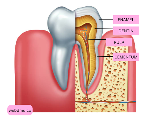
If you need a cheat sheet on Tooth Anatomy, here is an illustration that could help you out:

The two major divisions of the tooth
The tooth is divided into two major anatomical divisions: the crown and the root.
1st Division: The crown

The crown of a tooth refers to the visible, exposed part of the tooth that is covered by enamel. It serves several important functions:
Chewing and Grinding: The crown is primarily responsible for the mastication (chewing) process. Its shape, size, and surface texture are designed to facilitate the grinding and breakdown of food into smaller, more easily digestible pieces.
Protection: The crown acts as a protective covering for the underlying tooth structures, including the dentin, pulp, and root. The hard enamel layer shields these sensitive inner layers from mechanical forces, temperature changes, and chemical irritants.
Sensation: The crown is also involved in the transmission of sensations related to temperature, pressure, and texture. The enamel and dentin contain nerve endings that relay sensory information to the brain, helping us perceive and respond to different stimuli in the mouth.
Speech and Articulation: The shape and position of the crown, particularly in the anterior (front) teeth, contribute to proper speech and pronunciation. The interaction between the tongue, lips, and teeth is crucial for forming sounds and words accurately.
Aesthetics: The crown is an essential part of the smile and plays a significant role in facial aesthetics. Its appearance, color, and alignment influence the overall esthetics of a person’s smile and can impact self-confidence and social interactions.
When dentists restore or improve the crown esthetically, they may utilize various procedures such as dental fillings, dental crowns, veneers, or other cosmetic treatments to enhance the tooth’s appearance and function.
If we go further inside of the crown, we will then see that it has different layers. These hard tissue layers have different forms and functions. These layers are the following:
Enamel
In studying tooth anatomy, we will discuss the outermost layer and the hardest and most mineralized substance in the human body- the enamel.
The enamel is a hard tissue that encapsulates the crown of the tooth. What you have to remember about enamel is that aside from being the hardest substance in the body, it is composed of 95% inorganic material (primarily hydroxyapatite crystals) and only 5% organic material. This fact will come in handy when you start restoring the tooth.
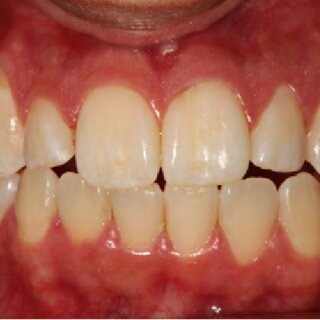
You should also note that the color of the enamel is not white. If you are indulged in the world of influencers and celebrities, you might think that you would also want those shiny white teeth. However, most of these individuals have veneers, which makes their teeth appear that way.
The real color of the enamel is grayish-white, and it is actually translucent. This is the reason behind the fact that some people would normally have yellow teeth compared to others. In essence, the enamel might be the outermost layer of the tooth, but the color of the dentin on the inside is also seen on the outside due to enamel’s translucency.
Here are the main functions of the enamel:
Protection:
Enamel provides a protective barrier for the underlying tooth structures, including the dentin and pulp. It shields the tooth from external forces, such as chewing and biting, and protects against physical and chemical damage.
Decay Resistance:
Enamel is highly resistant to acid and bacterial attacks, making it crucial in preventing tooth decay. Its dense and mineralized structure acts as a barrier, reducing the risk of cavities and erosion caused by acids produced by oral bacteria and dietary factors.
Sensitivity Reduction:
Enamel helps to reduce tooth sensitivity by covering and protecting the underlying dentin, which contains nerve endings. It acts as a barrier against temperature changes, sweet or acidic foods, and other stimuli that can trigger sensitivity.
Ethetics:
Enamel is responsible for the natural white color of teeth. Its translucent nature allows light to pass through and reflect off the underlying dentin, contributing to the appearance of a healthy and attractive smile.
Bite and Chewing Surface:
Enamel provides a smooth and durable surface for biting and chewing food. Its hardness and wear resistance allows for efficient mastication without significant wear or damage to the tooth.
It’s important to note that enamel, unlike other parts of our body, is irreplaceable. Therefore, maintaining good oral hygiene practices, such as regular brushing, flossing, and dental check-ups, is essential for preserving the health and integrity of enamel.
Dentin
The second layer of the crown is called the dentin, and compared to enamel, this layer is significantly softer; with only 70% inorganic content.
The interesting thing about dentin is that it contains microscopic tubules that extend from the inner layer of the dentin (near the pulp) to the outer layer, where they are exposed to the oral cavity. These tubules contain fluid and nerve endings, making dentin more sensitive to external stimuli, such as temperature, pressure, and acidic or sweet foods, compared to enamel.
Dentin is formed by specialized cells called odontoblasts, which are located in the pulp chamber. Odontoblasts produce and deposit dentin throughout life as a response to stimuli such as tooth decay, trauma, or dental procedures like cavity preparations.
Here are the main functions of the dentin:
Protection of Pulp: Dentin acts as a protective layer for the dental pulp, which is the soft tissue containing nerves and blood vessels at the center of the tooth. It acts as a barrier against irritants and helps to insulate the pulp from temperature changes and external threats.
Role in Tooth Color: Dentin plays a significant role in determining the color of a tooth. While enamel is translucent, dentin has a yellowish hue. The thickness and translucency of enamel influence how much dentin color is visible, contributing to the overall tooth color.
Secondary Dentin Formation: Dentin has the ability to regenerate and form new layers in response to various stimuli. When the tooth is exposed to factors like tooth decay, trauma, or dental procedures, specialized cells called odontoblasts produce secondary dentin to protect the pulp and restore the tooth’s integrity.
Pulp
The pulp is the soft, innermost part of a tooth that is located in the pulp chamber and root canals. It consists of connective tissue, blood vessels, nerves, and cells. This portion of the tooth supplies the blood and maintains the vitality of the tooth.
The main functions of the pulp include:
Nutrient Supply: The pulp contains a network of blood vessels that supply essential nutrients and oxygen to the tooth. These blood vessels nourish the cells within the pulp, ensuring their vitality and proper functioning.
Sensation and Innervation: The pulp contains a dense network of nerves, which make it highly sensitive to various stimuli. The sensory nerve fibers in the pulp allow us to perceive sensations such as temperature, pressure, and pain. The nerves in the pulp transmit signals to the brain, enabling us to respond to different stimuli in the oral cavity.
Defense and Repair: The cells within the pulp, including immune cells, play a role in the defense and repair mechanisms of the tooth. They help fight against bacteria and other pathogens that may invade the pulp and initiate the healing process when the tooth is damaged or injured.
Development and Tooth Maturation: During tooth development, the pulp plays a crucial role in the formation and maturation of the tooth. It provides the necessary nutrients and signaling molecules for the proper growth and differentiation of dental tissues, including enamel, dentin, and cementum.
When the pulp becomes infected, injured, or damaged, it may require treatment such as root canal therapy. Root canal treatment involves removing the infected or diseased pulp and filling the space with an inert material to save the tooth and alleviate pain or discomfort.
Now that we have discussed the crown and its parts, we can move on and discuss about the root.
2nd Division: The root
The root, on the other hand, is the part of the tooth anatomy that is located below the gum line and is not visible in the oral cavity. It serves several important functions:
Anchoring and Support
This portion of the tooth anchors it securely within the jawbone, providing stability and support. The roots extend into the alveolar bone of the jaw and are held in place by the periodontal ligament, which acts as a shock absorber and allows for slight movement during chewing.
Absorption of Forces
During biting and chewing, the forces generated are transmitted through the crown and into the root, which distributes the pressure evenly across the supporting bone. The root structure helps to absorb and dissipate these forces, protecting the surrounding tissues and preventing damage.
Nourishment
Blood vessels and nerves enter the tooth through small openings at the apex (tip) of the root. These blood vessels supply vital nutrients and oxygen to the pulp, which is located in the central chamber of the tooth and contains nerves and blood vessels. The root facilitates the exchange of nutrients and waste products between the tooth and surrounding tissues.
Sensation
Similar to the crown, the root contains nerve endings that transmit sensory information, allowing us to perceive sensations such as temperature, pressure, and pain. These nerve fibers extend from the pulp chamber within the crown down into the root.
Attachment of Supporting Structures
The root of the tooth serves as an attachment point for the periodontal ligament, which connects the tooth to the surrounding bone and helps maintain its position within the dental arch. The periodontal ligament also plays a crucial role in the transmission of forces during biting and chewing.
Just like enamel, the root is also comprised of different hard and soft tissues that are layered to encapsulate the pulp inside of it. If we dissect the root, we will be seeing these parts:
Cementum
The concept of cementum is not as common as the other parts of tooth anatomy like enamel and dentin. However, at the root part of the tooth, you will now see the cementum
Cementum is a specialized connective tissue that covers the root surface of a tooth. It plays a crucial role in anchoring the tooth to the surrounding bone within the dental socket. Here are some of the things that you have to know about cementum:
Attachment and Support
Cementum serves as a medium for attaching the periodontal ligament fibers to the tooth root. The periodontal ligament connects the tooth to the alveolar bone, providing support and stability. The attachment between the cementum and the periodontal ligament helps resist the forces generated during biting and chewing.
Protection of Dentin
Cementum covers the dentin that is exposed on the root surface. It acts as a protective layer, shielding the underlying dentin from external influences, such as mechanical and chemical damage. Cementum helps to maintain the integrity and health of the root structure.
Compensation for Occlusal Wear
As we age and engage in normal chewing activities, the occlusal (biting) surfaces of teeth may wear down. Cementum can continue to be deposited on the root surfaces throughout life, compensating for the loss of tooth structure and maintaining proper tooth length.
Repair and Regeneration
Cementum has limited regenerative capacity and can repair itself to some extent. When the root surface is damaged or exposed, cementoblasts, which are specialized cells, can produce new cementum to restore the protective layer.
Composition
Cementum is primarily composed of collagen fibers and a mineralized matrix. The mineral content of cementum is lower than that of dentin and enamel, making it softer. It has a slightly yellowish color.
Cementum is closely related to the health of the periodontal tissues and plays a crucial role in maintaining tooth stability and supporting proper oral function. Regular dental care, including professional cleanings and periodontal maintenance, is important to preserve the health and integrity of cementum and prevent gum diseases such as periodontitis.