
Your first year of dental school means getting to know the basics and memorizing them like the back of your hand. This means that you have to familiarize yourself with not just the anatomy and functions of your teeth, but with the structures that surround it as well, like the periodontium.
In this article, we will be talking about all the information that you know about periodontium. What it looks like, what comprises it, and the functions that it serves in our oral cavity. So make sure to grab your pens, papers, and drawing materials, and let’s get into it.
What is periodontium?
From the root word peri which means surround and odontos which means tooth, the periodontium refers to the soft and hard tissues and structures that surround the individual teeth in our oral cavity.
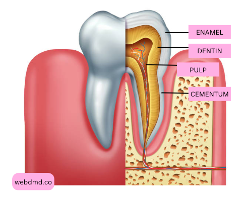
The periodontium includes the gingiva (gums), periodontal ligament, cementum, and alveolar bone. These components work together to provide stability, protection, and nourishment to the teeth. The periodontium plays a crucial role in maintaining the health and integrity of the teeth and their supporting structures.
What is the periodontium also known as?
The periodontium is also known as the periodontal apparatus, but it essentially means the same thing. It still serves the same purpose and has the same parts.
What are the 3 parts of the periodontium?
Lindhe, et. al recorded that the periodontium is composed of the following tissues: the gingiva or the gums, the periodontal ligament, the root cementum, and the alveolar bone proper.
Gingiva
As part of the masticatory mucosa, the gingiva or the gums covers the alveolar processes (the bone which covers the roots of the teeth) and the cervical portion of the teeth in a clinically healthy mouth.
When we say masticatory, what this means is that it is primarily involved in chewing, which is the case with the gingiva.
The gingiva can be divided into three main types: the free gingiva, the interdental gingiva, and the attached gingiva.
Free Gingiva
The free gingiva is the visible part of the gum that surrounds the teeth. It forms a collar-like band around each tooth and is not directly attached to the underlying bone.
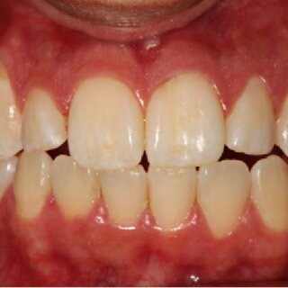
In a clinically healthy mouth, the free gingiva is positive parabolic in shape, coral pink in color, with firm consistency, and you can observe the presence of stippling. Upon complete eruption of the teeth, the free gingiva will normally be situated at around 1.5-2mm coronal to the CEJ.
In patients who suffer from periodontal diseases like gingivitis or periodontitis, the free gingiva appears to be erythematous in color, shiny, edematous, and not well-adapted to the alveolar process or the palate.
The free gingiva forms the gingival sulcus, a shallow groove between the tooth and the gum tissue.
Interdental Gingiva
The interdental gingiva, also known as the interdental papilla, is a specialized part of the gingiva that fills the space between adjacent teeth. Based on its name, it is located in the interproximal areas, which are the spaces between the teeth.
Some of the characteristics of the interdental gingiva include its pyramidal shape that heavily depends on the tooth position, the contact points of the adjacent teeth, and its anatomy. So for example, if you have a malposed tooth, your gingiva will more likely look blunted compared to that of the teeth that are properly aligned.
The interdental gingiva also provides support and stability to the adjacent teeth by filling the interproximal spaces. Aside from filling the gaps between the crowns of the teeth, it also helps in maintaining proper tooth alignment and prevents food impaction between the teeth.
Attached Gingiva
This is the part of the gingiva that is tightly bound to the underlying alveolar bone. It extends from the free gingiva to the mucogingival junction, which is the boundary between the oral mucosa and the attached gingiva.
The gingiva has several important functions:
Protection
The gingiva acts as a protective barrier, covering and shielding the underlying tissues and tooth roots from physical and microbial damage.
Support and Attachment
The gingiva supports the teeth by providing attachment to the underlying periodontal ligament and alveolar bone. It helps to maintain the teeth in their proper positions.
Sensation
The gingiva contains a rich supply of nerve endings, which contribute to the sense of touch and provide feedback during chewing.
Defense
The gingiva plays a role in the body’s defense mechanisms against oral bacteria and pathogens. It contains immune cells and produces antimicrobial substances to help prevent infections.
Periodontal Ligament
The periodontal ligament is usually difficult to visualize even in radiographs because we can’t actually see it there, but this is part of the periodontium that acts as an anchor which keeps the tooth inside the socket.
What are the three functions of the periodontal ligaments?
These are actually similar to the other ligaments in your body- it is a vascularized connective tissue that allows movements. In this case, the periodontal ligaments’ function includes allowing the needed mobility of our teeth, anchoring the teeth in the alveolar socket, and dissipating the forces that each tooth receives during function so that it will be absorbed by a much tougher part of our oral cavity- the alveolar bone.
In the periodontal ligament, we also see different bundles of collagen fibers that help serve its function. These fibers are the following:
- Alveolar Crest Fibers (ACF)
- Horizontal Fibers (HF)
- Oblique Fibers (OF)
- Apical Fibers (APF)
Cementum
Cementum is one of the most interesting parts of the tooth that you will have to come across with because its characteristics are like a marriage between the enamel and the dentin. It also is comprised of HAP crystals just like enamel, but it is capable of continuous deposition through the presence of cementoblasts, just like dentin.
Like the other layers of the teeth, cementum also has no blood or nerve coursing through its entirety, but it plays a very important role in your tooth. Did you know that the cementum is the only portion where the periodontal ligament fibers attach?
It is important that enamel terminates at around the CEJ and slowly transits into the cementum because these periodontal ligament fibers could not adhere to the enamel. If the entirety of our tooth is covered with enamel, then it will not be anchored in the alveolar bone.
Alveolar Bone Proper
The bone that houses each tooth is the alveolar bone proper. They are also called the alveolar processes or the alveolar sockets. As a dental student, I always think of it as the different slots that would fit each tooth perfectly. The alveolar bone or the alveolar process is present in both the maxilla and the mandible to support the teeth.
Here are some key features of the alveolar bone proper:
Socket Shape: The alveolar bone proper has a socket-like shape that is specifically designed to accommodate the roots of each tooth. It forms a precise fit around the tooth, providing stability and support.
Lamina Dura: The alveolar bone proper has a dense layer of compact bone called the lamina dura, which forms the inner lining of the tooth socket. It appears as a radiopaque line on dental X-rays and indicates a healthy periodontium.
Trabecular Bone: The alveolar bone proper also contains trabecular or cancellous bone, which fills the spaces between the compact bone and provides structural support.
For the functions of the alveolar bone, here are the things that you have to remember:
Tooth Support: The primary function of the alveolar bone proper is to provide support and anchorage for the teeth. It holds the teeth in their proper positions within the dental arches.
Resorption and Remodeling: The alveolar bone is dynamic and undergoes constant remodeling in response to functional forces. It can undergo resorption or deposition of bone depending on the forces applied to the teeth.
Periodontal Ligament Attachment: The alveolar bone proper provides attachment sites for the periodontal ligament fibers, which connect the tooth root to the surrounding bone. This attachment helps maintain tooth stability and allows for slight tooth movement during function.
To sum it all up
Understanding the importance of the periodontium and taking proactive steps to care for it can help prevent periodontal diseases and maintain optimal oral health.